Bone grafting
VIP bone grafting
Accelerated rehabilitation (up to three times faster than other clinics)
Treatment under conscious sedation without pain or stress
Bone grafting: What is it?
Bone grafting is a surgical procedure aimed to increase bone volume before placing a dental implant. The thickness of the bone at the implant site is very important. Insufficient bone volume could prevent the artificial root from being firmly attached. Failure to perform bone grafting and implant placement increases the risk of implant rejection.
Bone grafting cost
Does implantation require bone grafting?
Every clinical case is different. Even if external signs of bone atrophy are present (receding lip, sagging chin, etc.), the implantologist will decide whether bone grafting is necessary only after taking a CT scan.
Patients who have been missing teeth for a long time or suffer from inflammation (periodontitis) are at risk. There stands a high probability that their CT scan will reveal clear signs of severe bone loss.
However, I use innovative methods to place dental implants as soon as possible, which allows me to avoid bone grafting (in most cases). Make an appointment for a consultation. After careful examination, I will inform you whether teeth restoration with bone grafts is the optimal treatment option in your case.

Types of bone grafts in dentistry
Depending on the clinical situation, Tetri’s Smile uses one of the following types of bone grafts.
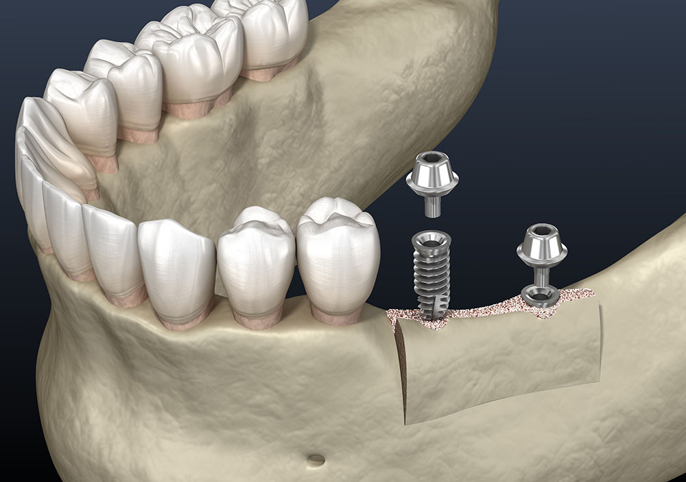
Alveolar ridge splitting
Indication: Horizontal bone atrophy, insufficient width of the alveolar process.
Performed at the same time as an implant is placed. The surgeon uses a piezotome (ultrasonic scalpel) to cut the alveolar ridge in the middle, screws in the implant, and fills the area around the implant with bone remnants. The site is covered with a protective membrane.
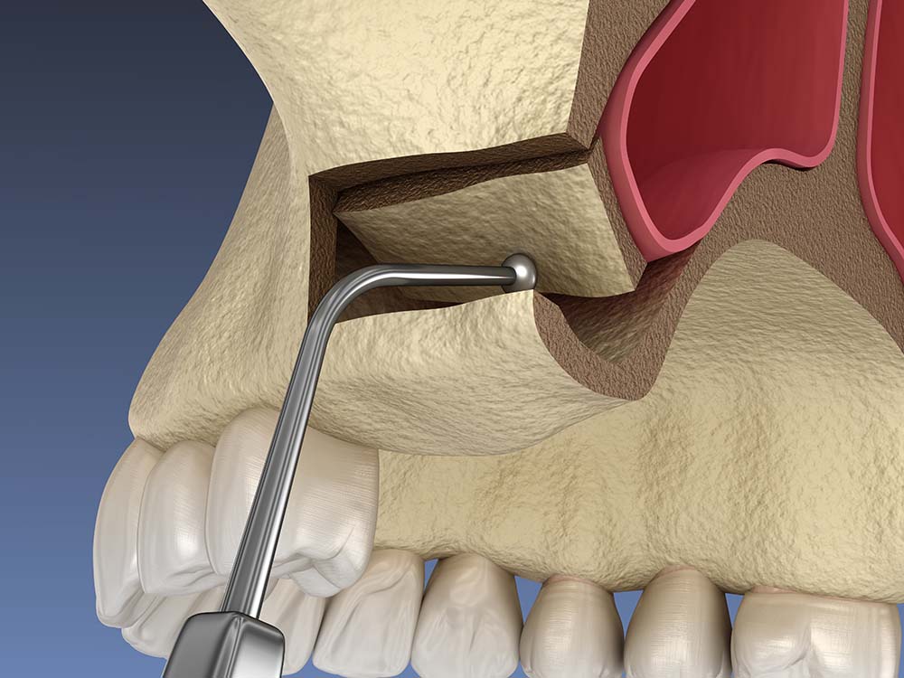
Indication: Vertical atrophy of the upper jaw, high risk of damage to the maxillary sinus.
Performed by creating lateral access to the sinus and elevating the sinus floor. The created space is filled with bone remnants. A protective membrane closes the incision and sutures are placed.
Bone block grafting
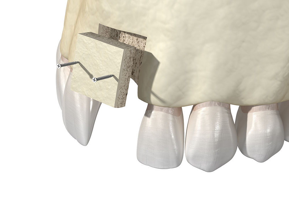
Indication: Bone loss in both width and height.
The bone block is taken from the patient (autogenic block sampling according to the method of Professor Kuri)
The implantologist screws the bone block to the jawbone and covers it with a protective collagen membrane.
Examples of bone grafting
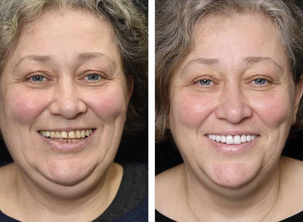
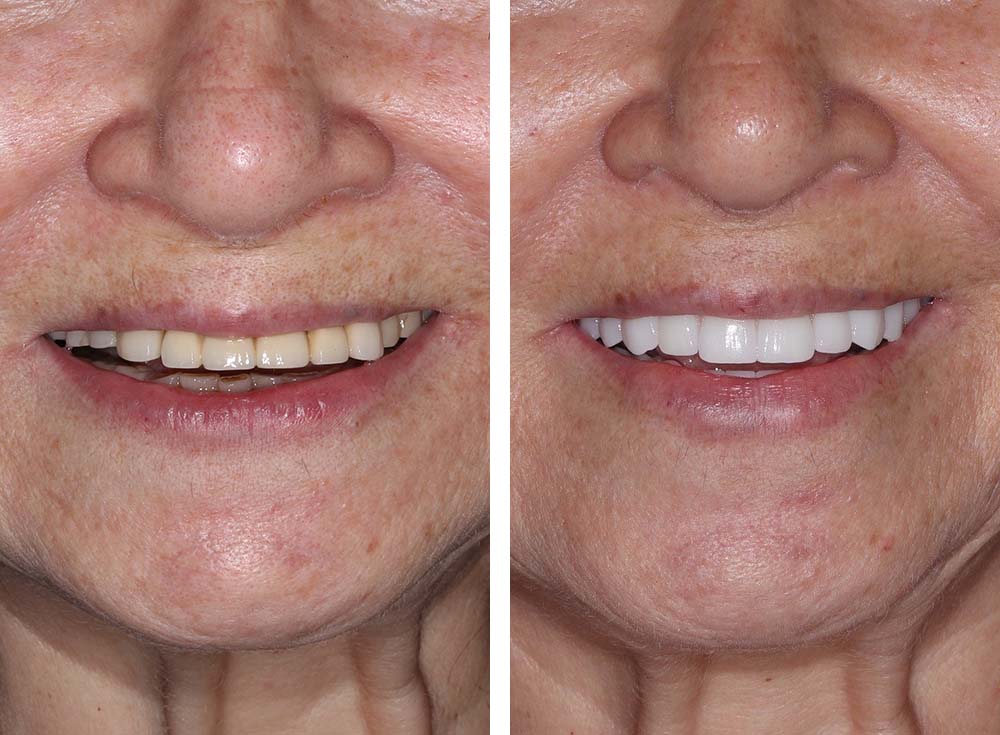
This patient came to our clinic complaining of missing several lower teeth and dissatisfaction with the aesthetics of her smile. A computed tomography scan revealed significant bone loss.
Dr. Tetri performed bone block grafting. When it took root, the implants were placed. After osseointegration of the implants, the doctor placed ceramic crowns on them. The front teeth in the smile area were restored with porcelain veneers.
This patient came to Tetri’s Smile with very loose implants and bridges that were incorrectly placed in another clinic, and suffered from a severe loss in bone volume.
Four implants in the lower jaw were removed, and bone grafting was performed.
The teeth in the upper jaw were extracted, for they could not be saved due to the advancement of periodontitis. Sinus elevation was performed, and seven implants with a full fixed overdenture were placed (all on x).
Preparing for bone grafting
Examination
The dentist visually analyzes the oral cavity and examines the patient’s medical history. If necessary, the patient is referred for a series of tests to rule out contraindications.
It is recommended to stop smoking and taking blood thinning medications at least one week before the bone grafting procedure.
CT scan
The dentist determines bone biotype based on CT scan results and assesses the degree of bone loss, calculating current height and width. The location of anatomically important structures such as maxillary sinuses, vessels, and nerves is determined. The presence of hidden neoplasms/cysts is excluded.
At Tetri’s Smile, the diagnosis is made using an ultra-precise Morita Veraviewepocs 3D CT scanner. We determine the height/width of the bone as accurately as possible. The position of sinuses, nerves and blood vessels are clearly visualized.
Oral cavity preparation
Before bone grafting, the dentist cleans the teeth of plaque and tartar, treats decayed teeth, and eliminates soft tissue inflammation.
Stages of bone grafting at Tetri’s Smile
Examination
The implantologist visually examines the oral cavity, and performs a CT scan. The data obtained is carefully analyzed to rule out possible contraindications to surgery. The optimal method of bone grafting is chosen.
Anesthesia
The implantologist administers an anesthetic to numb the surgical area. In combination with local anesthesia, it puts the patient into a medicated sleep (oral sedation).
Bone grafting
The implantologist performs an osteoplasty according to the selected technique, depending on the clinical picture.
Implant placement
If bone quality permits, the implant is placed immediately. If an anterior tooth is restored, a permanent crown is placed immediately. With a molar, a temporary crown is placed first, and after 3-5 months (the period of implant-bone fusion) it is replaced by a permanent one. In the case of full jaw implantation, the implants are loaded with a permanent overdenture on the same day.
You can go home immediately after surgery. Mild pain and swelling for the first 2-3 days after surgery is normal. Usually, by the 5th-7th day, the pain and swelling go away, and the patient feels no discomfort.
The bone block will fully take root 3-7 months after transplantation (depending on its size, individual characteristics of the body, and etc.).
Yes, but you must exclude spicy, sour, cold, hot, hard foods from your diet for the first 5-7 days post procedure.
In the first 7-10 days following the procedure, the following is not recommended: chewing on the operated side, smoking, alcohol consumption, sauna, flying.
During the first few days, it is important to brush your teeth carefully so as not to traumatize the postoperative area. It is recommended to use a toothbrush with soft bristles. You should not actively rinse your mouth with an antiseptic. Only oral baths are allowed.

My patients recover three times faster after bone grafting. First, I use the piezotome (ultrasonic scalpel), a gentle technique for working with bone tissue. Working with the piezotome does not traumatize soft tissues, vessels, and nerves. This results in less pain and bleeding after surgery, when compared to traditional bone grafting. Second, I use platelet-rich plasma and fibrin, stem cells, and other innovative developments to accelerate the rehabilitation process.
It is better to have a board-certified periodontist or oral surgeon perform the bone grafting procedure, in contrast to a general dentist.
Local anesthesia eliminates pain during bone grafting. At the patient’s request, sedation is used in combination with anesthesia, making the procedure stress- and worry-free.

